The Future of Eye Diagnostics
In an Intelligent World
Discover the latest diagnostic machines and intelligent tools shaping modern orthoptics and vision care.
Learn More


CF X-LINKER
CF X-LINKER
The CF X-LINKER® is an ophthalmic medical device for corneal cross-linking (CXL), manufactured by Iromed Group S.r.l. (Rome, Italy). It is used primarily in the treatment of keratoconus and other corneal ectasias.
The CF X-LINKER® is an ophthalmic medical device for corneal cross-linking (CXL), manufactured by Iromed Group S.r.l. (Rome, Italy). It is used primarily in the treatment of keratoconus and other corneal ectasias.

Yalkaid (YG-100K) SS-OCT:
Yalkaid (YG-100K) SS-OCT:
Swept-source OCT with full-range imaging capability, offering 12mm posterior scan depth and 15mm anterior scan depth with sub-6 micron optical resolution, utilizing 1060nm wavelength technology for improved penetration through cataracts and vitreous opacities
Swept-source OCT with full-range imaging capability, offering 12mm posterior scan depth and 15mm anterior scan depth with sub-6 micron optical resolution, utilizing 1060nm wavelength technology for improved penetration through cataracts and vitreous opacities
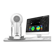
Scansys TA517 3D Anterior Segment Analyzer
Scansys TA517 3D Anterior Segment Analyzer
Scheimpflug-based anterior segment analyzer capturing 28-100 images (up to 384,000 data points) with 8th-order wavefront analysis, AI-powered keratoconus detection, and integrated calculators for refractive surgery, cataract planning, and contact lens fitting.
Scheimpflug-based anterior segment analyzer capturing 28-100 images (up to 384,000 data points) with 8th-order wavefront analysis, AI-powered keratoconus detection, and integrated calculators for refractive surgery, cataract planning, and contact lens fitting.
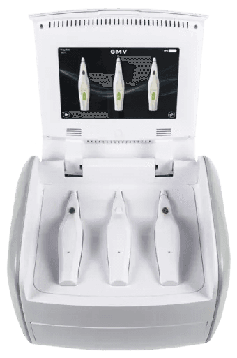
Plexr Plus
Plexr Plus
Plexr Plus is a patented micro-plasma device manufactured by GMV in Italy that generates controlled electric arcs through ionization of atmospheric gases.
Plexr Plus is a patented micro-plasma device manufactured by GMV in Italy that generates controlled electric arcs through ionization of atmospheric gases.
DREAM OCT - IntaLight
DREAM OCT - IntaLight
The DREAM OCT (Deep | Rapid | Extensive | Accurate | Multimodal) represents Intalight's advanced swept-source OCT system designed for comprehensive whole-eye imaging. Currently pending FDA approval and not yet available in the US market, this device combines multiple imaging modalities into a single platform for anterior and posterior segment evaluation.
The DREAM OCT (Deep | Rapid | Extensive | Accurate | Multimodal) represents Intalight's advanced swept-source OCT system designed for comprehensive whole-eye imaging. Currently pending FDA approval and not yet available in the US market, this device combines multiple imaging modalities into a single platform for anterior and posterior segment evaluation.
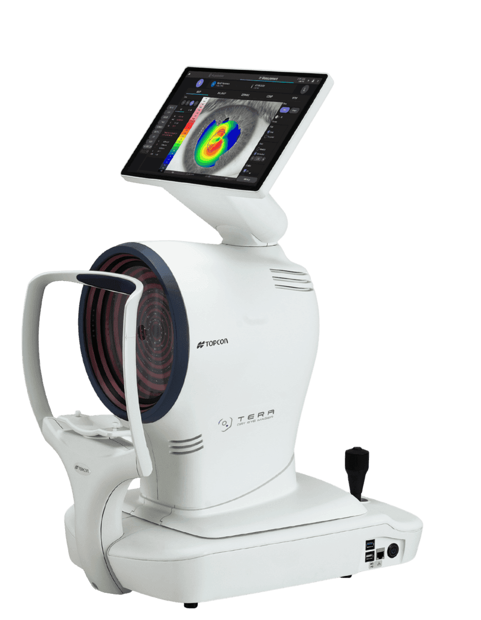
TERA Dry Eye Imager
TERA Dry Eye Imager
A comprehensive automated diagnostic system specifically designed for dry eye disease diagnosis and management.
A comprehensive automated diagnostic system specifically designed for dry eye disease diagnosis and management.
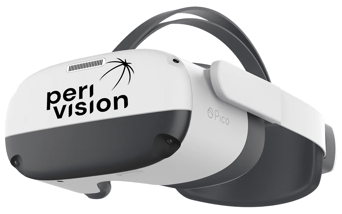
PeriVision VisionOne
PeriVision VisionOne
PeriVision VisionOne™ is a VR-based visual field testing platform for glaucoma diagnosis and monitoring.
PeriVision VisionOne™ is a VR-based visual field testing platform for glaucoma diagnosis and monitoring.
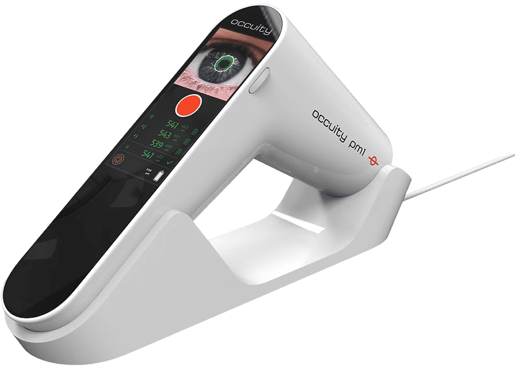
Occuity PM1 Pachymeter
Occuity PM1 Pachymeter
a device used to measure corneal thickness (specifically Central Corneal Thickness or CCT).
a device used to measure corneal thickness (specifically Central Corneal Thickness or CCT).
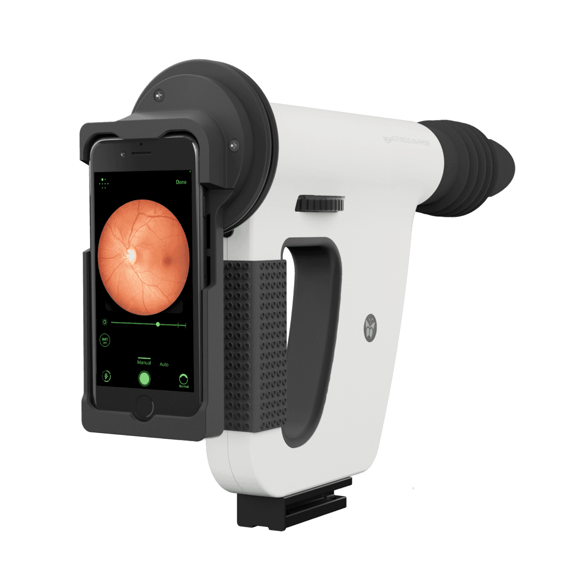
FOP NM-10 | Hand Held Fundus Camera
FOP NM-10 | Hand Held Fundus Camera
Remidio FOP (Fundus on Phone) is a handheld, portable retinal imaging device.
Remidio FOP (Fundus on Phone) is a handheld, portable retinal imaging device.

EmAA Pro 2
EmAA Pro 2
EMAA Pro 2 is an immersive software that places patients in a 3D virtual environment to analyze binocular vision disorders and perform rehabilitation, with objective eye-tracking measurements ensuring high accuracy.
EMAA Pro 2 is an immersive software that places patients in a 3D virtual environment to analyze binocular vision disorders and perform rehabilitation, with objective eye-tracking measurements ensuring high accuracy.

Integre® Pro Laser
Integre® Pro Laser
The system is designed for retinal photocoagulation procedures including focal laser treatment in the macular area and panretinal photocoagulation (PRP) in the periphery for various retinal pathologies.
The system is designed for retinal photocoagulation procedures including focal laser treatment in the macular area and panretinal photocoagulation (PRP) in the periphery for various retinal pathologies.

OCTOPUS 600 PERIMETER
OCTOPUS 600 PERIMETER
The Octopus 600 is an automated static perimeter manufactured by Haag-Streit Diagnostics, designed for visual field testing
The Octopus 600 is an automated static perimeter manufactured by Haag-Streit Diagnostics, designed for visual field testing

EM-4000 Tomey
EM-4000 Tomey
The Tomey EM-4000 is a high-speed specular microscope with LED illumination and automated CCD imaging, delivering endothelial analysis in 4 seconds with integrated pachymetry. Features proprietary "Core method" cell counting and auto-alignment with 16-image capture for accurate endothelial cell density and morphology assessment.
The Tomey EM-4000 is a high-speed specular microscope with LED illumination and automated CCD imaging, delivering endothelial analysis in 4 seconds with integrated pachymetry. Features proprietary "Core method" cell counting and auto-alignment with 16-image capture for accurate endothelial cell density and morphology assessment.

CASIA 2
CASIA 2
The Tomey CASIA 2 is a high-resolution anterior segment OCT system utilizing swept-source technology at 1310 nm wavelength, delivering 50,000 A-scans/second with 10 μm axial resolution across a 16 mm imaging diameter.
The Tomey CASIA 2 is a high-resolution anterior segment OCT system utilizing swept-source technology at 1310 nm wavelength, delivering 50,000 A-scans/second with 10 μm axial resolution across a 16 mm imaging diameter.

Optos Daytona
Optos Daytona
The Daytona represents the entry-level platform in Optos' ultra-widefield portfolio, providing essential UWF capabilities for practices requiring comprehensive retinal screening without the complexity of multi-modal advanced imaging systems.
The Daytona represents the entry-level platform in Optos' ultra-widefield portfolio, providing essential UWF capabilities for practices requiring comprehensive retinal screening without the complexity of multi-modal advanced imaging systems.

Optos California
Optos California
Confocal Scanning Laser Ophthalmoscopy (cSLO) Platform
Confocal Scanning Laser Ophthalmoscopy (cSLO) Platform

LSFG NAVI
LSFG NAVI
The LSFG NAVI is a non-invasive ophthalmic imaging device manufactured by Softcare Co., Ltd. (Japan) that uses laser speckle flowgraphy technology to quantitatively assess ocular blood flow in real-time.
The LSFG NAVI is a non-invasive ophthalmic imaging device manufactured by Softcare Co., Ltd. (Japan) that uses laser speckle flowgraphy technology to quantitatively assess ocular blood flow in real-time.

Optos MonacoPro
Optos MonacoPro
The MonacoPro is the only ultra-widefield retinal imaging device with integrated SD-OCT, producing a 200° single-shot optomap image in less than ½ second while providing cross-sectional 40° OCT views of retinal structures. Launched in February 2025, MonacoPro represents the next-generation ultra-widefield (UWF™) SLO and spectral domain retinal imaging solution building on the legacy of Monaco®.
The MonacoPro is the only ultra-widefield retinal imaging device with integrated SD-OCT, producing a 200° single-shot optomap image in less than ½ second while providing cross-sectional 40° OCT views of retinal structures. Launched in February 2025, MonacoPro represents the next-generation ultra-widefield (UWF™) SLO and spectral domain retinal imaging solution building on the legacy of Monaco®.

Triton™
Triton™
The Topcon DRI OCT Triton is a multi-modal swept-source OCT with a non-mydriatic color fundus camera, utilizing a 1,050 nm wavelength light source and a scanning speed of 100,000 A Scans/sec.
The Topcon DRI OCT Triton is a multi-modal swept-source OCT with a non-mydriatic color fundus camera, utilizing a 1,050 nm wavelength light source and a scanning speed of 100,000 A Scans/sec.

Maestro 2
Maestro 2
The Topcon Maestro2 is an automated multimodal imaging system combining 50,000 A-scans/sec Spectral Domain OCT with a non-mydriatic color fundus camera in a single, space-saving unit.
The Topcon Maestro2 is an automated multimodal imaging system combining 50,000 A-scans/sec Spectral Domain OCT with a non-mydriatic color fundus camera in a single, space-saving unit.
Visreed
One assessment at a time
VisReed 2025 © .
Visreed
One assessment at a time
VisReed 2025 © .