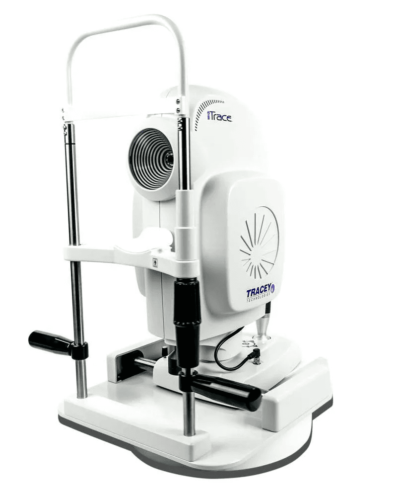
ITrace Wavefront Aberrometer
Abstract
The Tracey Technologies iTrace is a 5-in-1 ophthalmic diagnostic platform combining patented sequential ray-tracing aberrometry (256 points at 655/785nm wavelengths) with Placido-disk topography (24 rings, 8,640 points), autorefraction (±15D sphere/±10D cylinder), keratometry, and pupillometry (2.5-8mm range), uniquely separating corneal from internal aberrations and featuring proprietary DLI™/CPI™/QVI™ indices, marker-free Toric IOL planning/verification, and no-dilation post-op assessment capabilities.
DEVICE OVERVIEW
The iTrace is a 5-in-1 diagnostic ophthalmic system that combines wavefront aberrometry, corneal topography, autorefraction, keratometry, and pupillometry in a single integrated platform.
CORE TECHNOLOGY
Ray-Tracing Aberrometry
Technology Type: Sequential ray-tracing (forward aberration measurement)
Light Source: Near-infrared laser diode
Wavelength: 655 nm (topography) and 785 nm (ray tracing aberrometry)
Number of Measurement Points: 256 parallel laser beams projected sequentially
Measurement Principle: Measures where each light ray lands on the retina and quantifies light energy transfer
Unique Capability: Only aberrometer using forward ray-tracing (measures light going into the eye, not reflected back)
Corneal Topography
Technology: Placido disk-based system (Vista format by EyeSys Vision)
Number of Rings: 24 concentric rings
Measured/Analyzed Points: 8,640 data points
Corneal Coverage: 8 mm diameter on 42.5D sphere
Dioptric Range: 33.75 to 61.36 D
TECHNICAL SPECIFICATIONS
Refraction Measurements
Sphere Range: ±15.0 D
Cylinder Range: ±10.0 D
Measurement Accuracy: ±0.10 D
Reproducibility: ±0.10 D
Correlation with Manifest Refraction: T = 0.9925 (ranges -0.01 to -0.4 D)
Pupil Analysis
Pupil Scan Size Range: 2.5 mm to 8.0 mm diameter
Pupillometry: Automatic measurement under various lighting conditions
Day/Night Vision Analysis: Pupil size tracking for photopic and mesopic conditions
Keratometry
Measurement Type: Automated keratometry (flat and steep meridians)
Based On: Combined corneal topography and wavefront data
Applications: IOL calculations, toric lens planning, corneal power assessment
Aberration Analysis
Lower-Order Aberrations (LOA): Sphere, cylinder, defocus
Higher-Order Aberrations (HOA): Coma, trefoil, spherical aberration, secondary astigmatism, tetrafoil
Zernike Polynomial Analysis: Complete wavefront decomposition
Measurement Separation: Total, corneal, and internal (lenticular) aberrations analyzed separately
PHYSICAL SPECIFICATIONS
Footprint Dimensions: 13.0 inches (33.0 cm) × 17.0 inches (43.2 cm)
Weight: 27.4 lbs (12.4 kg / 20 kg depending on configuration)
Portability: Compact, space-saving design suitable for clinical settings
Capture Modes: Automatic and manual
CLINICAL ANALYSIS CAPABILITIES
Proprietary Indices (iTrace Prime Dashboard)
Dysfunctional Lens Index (DLI™)
Quantifies vision loss attributed to the crystalline lens
Scale: 0-10 (objective grading)
Used for cataract diagnosis and premium IOL candidacy
Corneal Performance Index (CPI™)
Quantifies vision loss attributed to the cornea
Scale: 0-10 (objective grading)
Identifies corneal aberrations and irregularities
Quality of Vision Index (QVI™)
Combined total eye visual quality metric
Sum of corneal and lenticular contributions
Comprehensive vision assessment tool
Tear Film Index (TFI)
Assesses ocular surface stability
Evaluates pre-operative tear film quality
Critical for refractive surgery screening
Visual Function Analysis
Retinal Spot Diagram (RSD): Graphical representation of total refraction, aberrations, and point spread function
Point Spread Function (PSF): Predicts retinal image quality
Modulation Transfer Function (MTF): Contrast sensitivity analysis
Multi-Zone Refraction Analysis: Refraction across different pupil zones
Wavefront Maps: Color-coded aberration maps (values in microns)
SPECIALIZED CLINICAL FEATURES
Toric IOL Planning & Verification
Toric Planner:
Integrated toric calculator using advanced iTrace keratometry
Portable eye image maps for precise intraoperative lens alignment
Uses natural iris landmarks for marker-free alignment
Optical alignment angle measurement
Toric Check (Post-Operative):
NO DILATION REQUIRED - 30-second examination
Determines if toric IOL is off-axis or off-power
Calculates predicted BCVA change with lens rotation
Provides rotation recommendations
Premium IOL Candidacy Assessment
Visual Axis Identification: Locates patient's visual axis relative to pupil center
Optical System Alignment: Determines multifocal IOL compatibility
Kappa Angle Measurement: Assesses distance between visual axis and limbus center
Pre-screening Tool: Predicts which patients will benefit from premium lenses
Patient Education Tools
Visual Simulation: Shows patients their actual vision vs. corrected vision
Snellen E Simulation: Demonstrates blurred vs. clear vision with/without correction
Toric vs. Standard IOL Comparison: Visual comparison for astigmatic patients
Objective Metrics: DLI, CPI, QVI communicated in patient-friendly format
DATA PROCESSING & OUTPUT
Acquisition & Analysis
Scan Time: Under 1 minute for complete profile
Data Processing: Point-by-point sequential analysis
Internal Optometer: Automatic patient alignment and accommodation control
Fogging Range: +7D to -5D (eliminates accommodation)
Fixation: Binocular and monocular open-field (reduces accommodation)
Integration & Connectivity
DICOM Compliance: Full integration (Version 6.3+)
Network Capability: Networkable system
Worklist Import: Managed worklist reduces data entry errors
Batch Processing: Batch saving and printing capabilities
Report Generation: Comprehensive, easy-to-interpret printable reports
Measurement Capabilities
Over-Spectacle Measurement: Can measure through spectacles
Post-Operative Analysis: Functions through IOLs, contact lenses, after LASIK
Highly Aberrated Eyes: Effective even with significant optical irregularities
Pseudophakia: Accurate measurements in patients with IOLs
Cataract Assessment: Can measure through early cataracts
CLINICAL APPLICATIONS
Refractive Surgery
Pre-operative screening and planning
LASIK/PRK candidate evaluation
Post-operative outcomes assessment
Identifying causes of post-operative visual complaints
Wavefront-guided treatment planning
Cataract Surgery
Dysfunctional Lens Syndrome diagnosis
Premium IOL candidate selection (multifocal, toric, accommodating)
IOL power calculation optimization
Toric IOL axis planning and verification
Post-operative lens position assessment
General Ophthalmology & Optometry
Differential diagnosis of vision complaints
Corneal disease monitoring (keratoconus, irregular astigmatism)
Contact lens fitting optimization
Second-opinion evaluations
Progression tracking of lens and corneal dysfunction
Research Applications
Clinical studies requiring precise optical measurements
Aberration analysis in various patient populations
IOL performance evaluation
Normal population reference databases
SOFTWARE VERSIONS & UPGRADES
Current Version: iTrace 7.0 (iTrace Prime)
Key Features:
Prime Dashboard (DLI, CPI, QVI indices)
Enhanced imaging quality
Improved wavefront capture process
Enhanced navigation between displays
Batch saving and printing
Toric Planner customization improvements
Tech Triage icon (quality control for technicians)
Exam Results display (validates scan quality)
Previous Major Versions:
Version 6.3: Added DICOM compliance
Version 6.1: Introduced Toric Check feature
Version 6.0: Introduced Dysfunctional Lens Index (DLI)
Version 4.0: Made diagnostic aberrometry accessible to all physicians
Upgrade Policy: Existing iTrace owners can upgrade software to access latest features
MEASUREMENT ACCURACY & VALIDATION
Clinical Studies
High concordance with manifest refraction (T = 0.9925)
More accurate than Hartmann-Shack aberrometers in post-refractive surgery eyes
Excellent repeatability for spherical aberration and multiple aberrometric coefficients
Intraclass correlation coefficients (ICC) > 0.96 for angle kappa measurements
Repeatability
Low within-subject standard deviation (Sw) for most measurements
Coefficient of variation (COV) better for lower-order aberrations
Repeatability affected by tear film quality (assess tear film before imaging)
Interdevice Comparisons
Comparable to Pentacam, OPD-Scan, and other aberrometers for specific parameters
Unique capability to separate corneal from internal aberrations
Different measurement principles may yield non-interchangeable Zernike coefficients
COMPETITIVE ADVANTAGES
Only Ray-Tracing Aberrometer: Measures forward aberrations (how eye actually uses light)
Corneal vs. Internal Separation: Only device that mathematically separates corneal from lenticular aberrations
Post-Operative Capability: Accurate measurements through IOLs, contact lenses, after refractive surgery
No Dilation Toric Check: Unique post-operative toric assessment without dilation
5-in-1 Integration: Eliminates need for multiple separate devices
Patient Education: Visual simulation capabilities for enhanced patient communication
Compact Footprint: Space-efficient design vs. multiple standalone devices
COMPATIBILITY & INTEGRATION
Compatible with major EMR/EHR systems (via DICOM)
Integrates with surgical planning software
Exports data for IOL calculation programs
Network-ready for multi-location practices
Standalone computer included (HP Panel PC with Intel i5 processor)
CLINICAL VALIDATION & ADOPTION
Used extensively in research institutions for clinical studies
Adopted by thousands of ophthalmology and optometry practices worldwide
Referenced in numerous peer-reviewed publications
Standard of care for practices performing premium cataract surgery
Endorsed by leading ophthalmologists for toric and multifocal IOL procedures