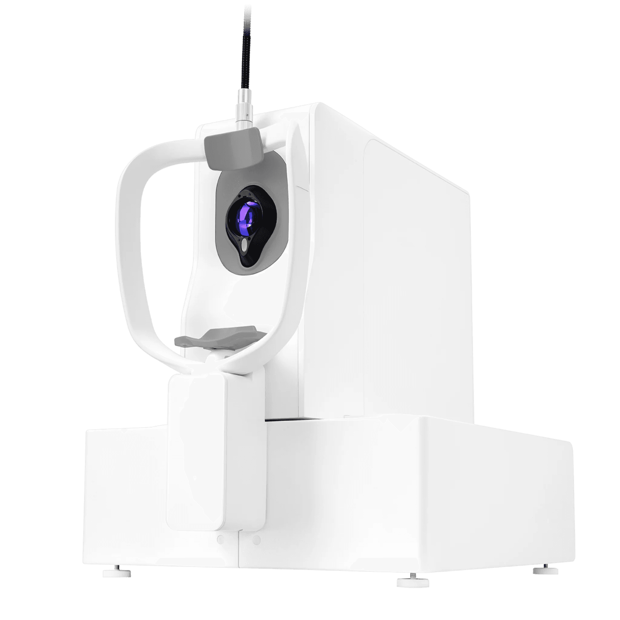
DREAM OCT - IntaLight
Abstract
The DREAM OCT (Deep | Rapid | Extensive | Accurate | Multimodal) represents Intalight's advanced swept-source OCT system designed for comprehensive whole-eye imaging. Currently pending FDA approval and not yet available in the US market, this device combines multiple imaging modalities into a single platform for anterior and posterior segment evaluation.
Technical Specifications
OCT Imaging System
Core Technology:
Methodology: Swept-Source OCT (SS-OCT)
Light Source: Swept-source tunable laser with center wavelength between 1030-1070 nm
Scan Speed: 200,000 A-scans per second (also available in 400,000 and 100,000 configurations depending on country registration)
Resolution & Depth:
Axial Resolution: ≤ 5.5 μm in tissue
Transverse Resolution: ≤ 15 μm in tissue
A-scan Depth: 12.0 mm in tissue
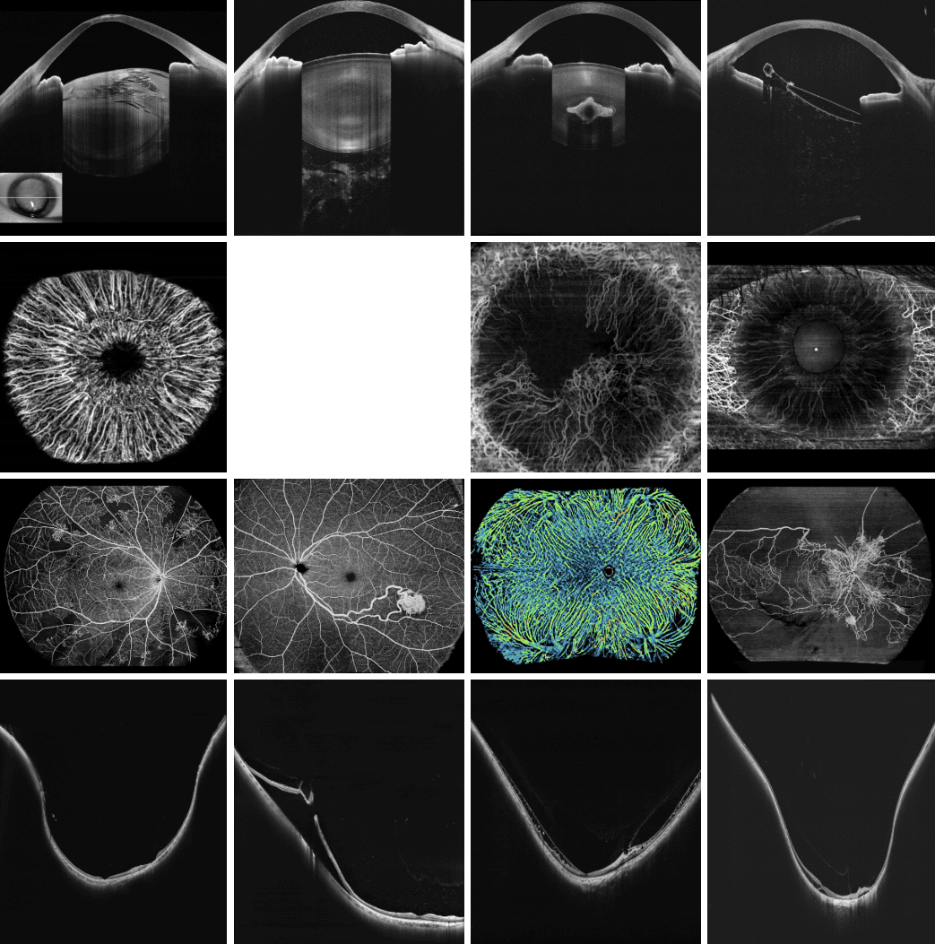
Fundus Imaging System
Methodology: Confocal Scanning Laser Ophthalmoscope (cSLO)
Light Source: Super Luminescent Diode (SLD), 810-850 nm wavelength
Additional Modalities
OCT Angiography (OCTA): Included for vascular imaging
Biometry: Integrated biometric measurements for surgical planning
Anterior Segment Imaging: Full anterior segment analysis capability
Key Technological Features
1. "All-in-One" Platform
The DREAM OCT consolidates multiple diagnostic functions into a single device:
Posterior segment OCT imaging
Anterior segment OCT
OCTA (vascular imaging)
Fundus photography via cSLO
Biometric measurements for cataract surgery planning
This integration eliminates the need for multiple separate devices, streamlining workflow and reducing equipment costs.
2. Ultra-High Speed Imaging
At 200,000 A-scans per second (with options up to 400,000 A-scans/sec in some regions), the DREAM OCT offers:
Reduced motion artifacts: Faster scanning minimizes eye movement during acquisition
Higher density sampling: More data points create more detailed images
Improved patient comfort: Shorter exam times reduce patient fatigue
Better repeatability: Quick scans improve test-retest reliability
3. Deep Penetration with Swept-Source Technology
The 1030-1070 nm wavelength range offers significant advantages:
Enhanced choroidal visualization: Longer wavelengths penetrate deeper through pigmented tissues
Reduced light scattering: Better image quality in eyes with media opacities (cataracts, vitreous hemorrhage)
Full-thickness retinal imaging: From internal limiting membrane to deep choroid in a single scan
Myopia research applications: Essential for studying sclera and posterior pole changes
4. PAR (Precision Anterior-Posterior Ratio) Technology
The device features sophisticated optical correction algorithms:
Distortion correction: Compensates for optical distortions in raw images
Refraction correction: Accounts for light refraction through different ocular media
Accurate biometry: Ensures precise measurements of axial length, corneal power, lens thickness, and anterior chamber depth
Clinical precision: Critical for IOL (intraocular lens) power calculations in cataract surgery
5. Extended Imaging Depth
With 12.0 mm imaging depth in tissue, the system can capture:
Complete anterior segment (cornea to lens)
Full posterior segment (retina to deep choroid)
Optic nerve head with surrounding structures
"Whole eye" visualization in comprehensive scans
Clinical Applications
Posterior Segment Diseases:
Age-related macular degeneration (AMD)
Diabetic retinopathy and macular edema
Glaucoma (retinal nerve fiber layer and ganglion cell analysis)
Choroidal disorders (choroidal osteoma, as referenced in published literature)
Myopia progression monitoring (particularly relevant given cited research on dietary fatty acids and myopia)
Anterior Segment Assessment:
Corneal pathology and dystrophies
Angle closure evaluation
Post-refractive surgery monitoring
Anterior chamber depth measurements
Surgical Planning:
Cataract surgery biometry
IOL power calculations
Refractive surgery screening
Glaucoma surgery planning
Vascular Imaging:
OCT Angiography for diabetic retinopathy
Macular telangiectasia
Retinal vein occlusions
Choroidal neovascularization in AMD
Research Applications
The device has been utilized in peer-reviewed research, including:
Choroidal Osteoma Study (Zhou et al., Frontiers in Oncology, 2021): Demonstrated the system's ability to visualize tumor vasculature using SS-OCTA, showing its utility in rare ocular tumors.
Myopia Research (Pan et al., PNAS, 2021): Used in groundbreaking research showing dietary omega-3 polyunsaturated fatty acids' protective effects against myopia, highlighting the device's research-grade imaging capabilities.
Competitive Advantages
Speed Comparison:
At 200,000-400,000 A-scans/second, DREAM OCT competes with high-end systems like:
Zeiss PLEX Elite (up to 200,000 A-scans/sec)
Topcon Triton (100,000 A-scans/sec)
Heidelberg Spectralis (85,000 A-scans/sec with standard module)
Swept-Source Benefits Over Spectral-Domain OCT:
Deeper penetration (essential for choroid imaging)
Less sensitivity roll-off with depth
Better performance through media opacities
Faster scan speeds possible
Superior for anterior segment imaging
Multimodal Integration:
Unlike single-purpose devices, the all-in-one design reduces:
Equipment footprint in clinic
Initial capital investment
Staff training requirements
Data management complexity
Limitations & Considerations
Regulatory Status:
Not FDA approved for use in the United States
Availability varies by country and regulatory approval status
Market Position:
Relatively new entrant competing against established brands (Zeiss, Heidelberg, Topcon, Optovue)
Limited clinical track record compared to legacy systems
May require building service/support network in new markets
Technical Considerations:
Swept-source systems generally more expensive than spectral-domain OCT
Longer wavelength may provide less contrast for inner retinal layers compared to shorter wavelengths
Requires regular maintenance and calibration like all precision optical instruments
Clinical Workflow Integration
Typical Use Case:
Patient positioning: Automated or manual alignment
Quick fundus preview: cSLO provides real-time imaging for targeting
Automated scanning: High-speed acquisition of volumetric data
PAR correction: Automatic post-processing for biometric accuracy
Multi-modal review: Clinician reviews OCT, OCTA, and fundus images in integrated software
Report generation: Comprehensive documentation for medical records
Time Efficiency:
Single-device workflow eliminates patient movement between instruments
High scan speeds reduce chair time
Automated analysis speeds diagnosis
Future Outlook
Technology Trends:
Increasing adoption of swept-source technology as prices decrease
Growing demand for integrated, multimodal platforms
AI integration for automated detection and analysis (likely future development)
Telemedicine compatibility for remote reading
Market Opportunities:
Practices seeking to consolidate equipment
High-volume clinics needing speed and efficiency
Research institutions requiring advanced imaging
Emerging markets where space and budget are constrained
Conclusion
The DREAM OCT represents a sophisticated, multimodal imaging platform that addresses the modern ophthalmic practice's need for comprehensive, efficient diagnostics. Its swept-source technology, ultra-high speed, deep penetration, and all-in-one design position it as a competitive option for practices seeking to future-proof their imaging capabilities. However, its current limitation in FDA approval restricts immediate US market adoption. Once regulatory clearance is obtained, it has potential to compete effectively in the premium OCT segment, particularly for practices valuing workflow integration and whole-eye imaging capabilities.
The published research utilizing this device demonstrates its research-grade quality, while its technical specifications suggest it can meet the demanding requirements of both clinical practice and investigative ophthalmology. As the ophthalmic imaging market continues to evolve toward integrated, AI-enhanced platforms, the DREAM OCT's foundation positions it well for future software enhancements and expanded clinical applications.