

Vantage Plus Digital - Keeler
From the manufacturer
Keeler: official product page
keelerglobal.com · opens in a new tab
Abstract
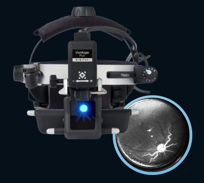
The Vantage Plus Digital is a head-mounted binocular indirect ophthalmoscope (BIO) from Keeler with a built-in 3.2 MP camera. It lets the examiner see the retina through the usual stereoscopic optics while the same view is displayed, recorded and saved as images and video in real time. Images are managed in Keeler's Kinexis software, with report generation and DICOM export. An optional fluorescein angiography (FA) upgrade kit adds a blue excitation light and yellow barrier filters, so FA can be documented through the BIO, including in the far periphery.
Core Technology Specifications
Instrument Type: Digital binocular indirect ophthalmoscope, headband design
Camera: 3.2 MP, Sony sensor technology; image size 2048 x 1536
Camera Interface: USB 3.0, for a high frame rate and smooth video
Camera Adjustment: ±7°, independent of the BIO optics
Illumination: LED, CRI 90
Working Distance: 30 to 65 cm (with a 20 D lens)
Depth of Field: Up to 100 mm
Pupil Size: 1 to 10 mm
Pupillary Distance: 48 to 76 mm; automatic stereopsis adjustment
Patch Size: 20, 40 or 60 mm (S / M / L)
Software: Keeler Kinexis (Windows 10/11, Intel i5 or higher, 8 GB RAM, 1920 x 1080 display, USB 3.0)
Optional Accessories: FA upgrade kit, foot switch; travel case included
Manufacturing: United Kingdom and United States
Anatomical Context: Viewing the Retina by Indirect Ophthalmoscopy
Indirect ophthalmoscopy views the fundus through a handheld condensing lens, typically 20 D, held in front of the patient's eye. The examiner sees an inverted, reversed, stereoscopic image with a wider field than direct ophthalmoscopy and a good view through media opacities. With the patient's eye rotated and scleral indentation, the examiner can reach the retinal periphery up to the ora serrata, which is hard to reach with most other methods.
The Vantage Plus Digital keeps this optical approach and adds a camera, so the findings that were previously only seen can now also be documented, compared and shown to the patient, the family or students.
How the System Works
- Optics: Keeler optics, aligned with laser alignment techniques, give a clear and sharp stereoscopic image. A customized optical design aims to keep the image stable during the examination
- Field and depth: The widened field of view covers more than an entire 20 D lens, and the depth of field of up to 100 mm accommodates changes in lens position and examination angle
- Camera: The 3.2 MP camera captures images and videos in real time. It can be adjusted by ±7° separately from the BIO optics, so the recorded view can be matched to what the examiner sees
- Capture and review: Kinexis stores recordings, from which still frames can be extracted at any point after the exam
- Controls: An adjustable focus knob adapts the device to different users, and an optional foot switch can trigger capture
Fluorescein Angiography Kit
The FA upgrade kit is the newest addition to the Vantage Plus Digital. Keeler describes it as an industry first in indirect retinal imaging.
- Excitation: A dedicated high-intensity blue excitation LED provides the light that makes the fluorescein dye glow
- Barrier filters: Precision yellow barrier filters let the emitted green-yellow fluorescence through and block the reflected blue light. Keeler says they give brighter, higher-contrast vascular detail than generic filters
- Peripheral reach: FA can be documented into the extreme retinal periphery, including during scleral indentation, without additional devices
- Footprint: Keeler states that it avoids the cost, complexity and space of traditional FA systems
- Availability: The Keeler global page says the FA kit is not available in the USA. Check availability in your country
Kinexis Software
- Purpose: A documentation suite for workflow management and ophthalmoscopy image capture
- Images and reports: Extracts still frames from recorded video and generates detailed reports
- DICOM: Exports data to DICOM, so findings can be shared with other systems
- Comparison and teaching: The brochure also lists side-by-side image comparison and live-streaming of examinations for teaching
Clinical Applications
- Documentation of the fundus and the retinal periphery during routine examination
- Documentation of peripheral findings such as tears, detachments or lesions, with scleral indentation
- Fluorescein angiography documentation with the FA kit, for example for vascular leakage and non-perfusion
- Pediatric retinal documentation, as reported by a user at Aravind-Coimbatore
- Teaching, using the live view and recorded images
- Referral and follow-up, by comparing images between visits
Who Can Use It
Ophthalmologists and retina specialists for fundus and FA documentation, residents and fellows in training, and optometrists with indirect ophthalmoscopy skills. Orthoptists and technicians can use the recorded images and Kinexis reports in clinic workflows, while FA itself involves intravenous dye and should be done under a clinician's supervision.
Comparison with a Standard BIO
| Feature | Standard BIO | Vantage Plus Digital |
|---|---|---|
| Recording | None | 3.2 MP images and video |
| Review | Memory and drawing | Kinexis, frames and reports |
| Teaching | Observer needs a teaching head | Live view and video |
| Data sharing | Manual | DICOM export |
| FA | Not possible | Optional FA kit |
Strengths and Limits
Strengths: A familiar BIO workflow with digital documentation, a widened field and deep depth of field, a camera that is adjustable independently, DICOM-ready software, and an optional FA kit that avoids a separate FA system.
Limits: It is wired by USB 3.0 to a computer. Image quality depends on the examiner's lens handling, pupil dilation and media clarity. A 3.2 MP camera gives less detail than dedicated widefield imaging systems. The FA kit is not available in the USA.
About Keeler
Keeler is a British ophthalmic instrument maker with a history of more than a century, known for ophthalmoscopes and slit lamps. The Vantage Plus Digital is made in the United Kingdom and the United States.
Limitations of Available Specifications
Note: The sources reviewed were the Keeler global product page and brochure, a distributor page and a retailer listing. Weight, battery or power details, the headset and lens specifications of the FA kit, the excitation wavelength, the image storage capacity, regulatory details by country and pricing were not available. A brochure lists dimensions of 35 x 23 x 22 cm, which probably refers to the packaged unit, but this was not stated. One retailer page for the older wireless Vantage Plus LED model was not used for the digital model. Contact Keeler or an authorized distributor for the full data sheet. This content supports clinical judgement and does not replace it.