Scansys TA517 3D Anterior Segment Analyzer
Abstract
Scheimpflug-based anterior segment analyzer capturing 28-100 images (up to 384,000 data points) with 8th-order wavefront analysis, AI-powered keratoconus detection, and integrated calculators for refractive surgery, cataract planning, and contact lens fitting.
Manufacturer: Shanghai MediWorks Precision Instruments Co., Ltd.
Technology: Rotating Scheimpflug imaging system for comprehensive anterior segment analysis
Core Imaging Technology
Imaging Method: Scheimpflug imaging system
Image Acquisition Options:
28 ultra-high-definition Scheimpflug images (107,520 data points)
60 ultra-high-definition Scheimpflug images (230,400 data points)
100 ultra-high-definition Scheimpflug images (384,000 data points)
Coverage Area: 16mm diameter of corneoscleral area (for scleral lens module with 60-image mode)
Measurement Capabilities
Corneal Analysis:
Central corneal thickness (CCT) and mid-peripheral corneal thickness (MPCT)
Corneal curvature measurements
Anterior and posterior corneal surface topography
Corneal diameter (White-to-White)
Eccentricity (ecc) values in different directions (nasal, temporal, inferior, superior, flat, steep) for diameter ranges 2-10mm
Sagittal height measurements
Corneal thickness spatial profile (CTSP)
Percentage thickness increase (TCI)

Anterior Chamber Analysis:
Anterior chamber depth (ACD)
Anterior chamber volume
Anterior chamber angle measurements
Angle opening distance (AOD)
Trabecular-iris space area (TISA)
Kappa angle and alpha angle
Lens Analysis:
Crystalline lens density analysis (cross-sectional and longitudinal sections via gray value calculation)
Corneal Refractive Power Measurements
SimK (simulated keratometry)
Total corneal power
True net power
Total corneal astigmatism
Ratio of posterior to anterior corneal surface curvature radius
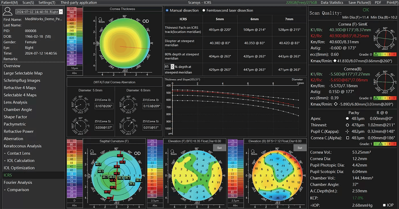
Aberrometry
Wavefront Analysis:
Up to 8th-order Zernike polynomial decomposition
Analyzed for: anterior corneal surface, posterior corneal surface, and total cornea
Maximum diameter: 12mm
Total corneal spherical aberration
Total corneal higher-order aberrations
Visual Quality Simulation:
Retinal imaging simulation
Point spread function (PSF)
Modulation transfer function (MTF)
Advanced Diagnostic Features
Keratoconus Detection:
Keratoconus index (KCI) for anterior and posterior surfaces
SVM (Support Vector Machine) classifier
Keratoconus probability (KCP) scoring
Keratoconus grading (Grade 0-IV)
Refractive 4 Maps analysis
Case reference database
Bilateral differential topography algorithm
Fourier Analysis: Decomposes corneal topography into:
Spherical component
Decentration component
Regular astigmatism component
Irregularity component
Built-in Calculators & Databases
IOL Calculation Formulas:
Pearl-DGS (optional)
Jin formula
SRK-T
Holladay 1
Hoffer Q
Age optimization for astigmatism
Age optimization for spherical aberration
Contact Lens Fitting:
Automatic calculation for orthokeratology lenses, soft contact lenses, and RGP lenses
3D/2D simulated fitting with fluorescence staining simulation
Customizable contact lens database
IOP Correction: 5 built-in intraocular pressure correction formulas based on corneal thickness
Database
Global population database: Approximately 3,000 cases (and growing)
Clinical Applications
Refractive surgery screening
Refractive cataract surgery planning
Contact lens fitting (including orthokeratology and scleral lenses)
Glaucoma screening
Intracorneal ring segment (ICRS) surgery planning
ICL (Implantable Collamer Lens) size recommendation and vault prediction/measurement