

RS-1 Glauvas - NIDEK
From the manufacturer
Nidek: official product page
nidek-intl.com · opens in a new tab
Abstract
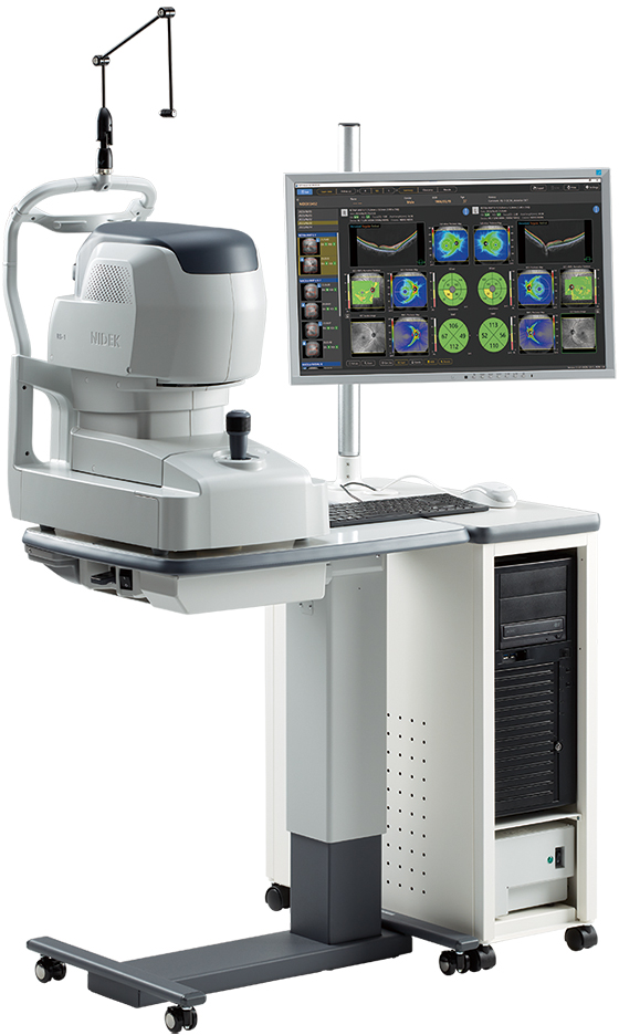
The RS-1 Glauvas is a spectral-domain optical coherence tomography (OCT) system from NIDEK, designed for glaucoma and retinal practice. It scans at up to 250,000 A-scans/s with an 880 nm SLD source and an SLO fundus image, and it can capture a single B-scan from the optic nerve head to the temporal vascular arcade. Its glaucoma tools include scan width correction and a long axial length normative database (Asian sample) for myopic eyes, deep-learning segmentation, and a Structural Normality Map. OCT angiography is optional. NIDEK announced the launch on 3 June 2024.
Core Technology Specifications
Instrument Type: Spectral-domain OCT with SLO fundus imaging
Light Source: SLD, 880 nm
Scan Speed: Up to 250,000 A-scans/s
Optical Resolution: Z 7 µm, X-Y 20 µm (the brochure table is jumbled and these values were matched by position, so they are not explicitly labeled as axial and transverse)
Scan Depth: 4.2 mm (one reseller page also lists a 9 mm depth, which conflicts with this)
Scan Width: Line up to 16.5 mm; map up to 15.0 x 12.0 mm
Image Averaging: Up to 120 images
Fundus Imaging: SLO, angle of view 53.3° x 53.3°
Minimum Pupil Diameter: 2.5 mm
Diopter Correction Range: −20 D to +20 D
Working Distance: 24.9 mm in fundus OCT mode; 20.0 mm in anterior mode
Fixation: Internal lamp green; external lamp red
Alignment: Automatic (X-Y-Z); the operator sets chinrest height, then selects Optimize and Capture
OCT Angiography (optional): 6 x 6, 9 x 9 and 12 x 12 mm scans; CODAA 2 algorithm (Complex OCT signal Difference Analysis Angiography 2)
Power: 100 to 240 V AC, 50/60 Hz; 220 VA for the main body
Dimensions and Weight: 332 (W) x 526 (D) x 586 (H) mm; 30.6 kg
Anatomical Context: Glaucoma and the Myopic Eye
Glaucoma OCT compares the thickness of the retinal nerve fiber layer (RNFL) and the ganglion cell complex (GCC) with a reference database. Highly myopic eyes are a known difficulty. Axial elongation changes the geometry of the retina and the optic disc, and a standard database built on mostly non-myopic eyes can flag healthy long eyes as abnormal. This is the problem the long axial length database and scan width correction are meant to address.
This paragraph is general clinical background and is not taken from the sources.
How the System Works
- Acquisition: A spectral-domain OCT with an 880 nm SLD acquires up to 250,000 A-scans/s. NIDEK says this shortens capture time and reduces fixation errors
- Fundus image: The SLO shows a 53.3° x 53.3° fundus image during capture
- Wide, deep scans: A single line scan of up to 16.5 mm can cover the optic nerve head and the temporal vascular arcade; NIDEK says the 4.2 mm depth captures the oblate shape of myopic eyes
- Axial length handling: Scan width correction. NIDEK's page says axial length is estimated from the OCT image, not measured directly, and is used for this correction
- Segmentation: Segmentation of 7 retinal layers, with deep-learning segmentation that NIDEK says reduces artifacts and false positives in the normative and thickness maps, including in eyes with opacities
- Options: Optional anterior segment kit for corneal thickness, corneal thickness map and angle measurement, and optional B-scan denoising software
Software Analysis and Normative Databases
The software list from the brochure includes: full retina thickness map, GCC thickness map, RNFL thickness map, percentile maps (RNFL, GCC+RNFL), Structural Normality Map, disc analysis and follow-up analysis.
| Database | Axial length | Age |
|---|---|---|
| Standard | Less than 26 mm | 20 to under 80 years |
| Long axial length (Asian sample) | 26 mm to less than 29 mm | 20 to under 60 years |
Both databases cover the macula map, disc map and retina map patterns. The ethnic composition of the standard database is not stated in the pages reviewed. The Structural Normality Map was developed with Kagoshima University, according to NIDEK's launch news.
Clinical Applications
- Structural glaucoma assessment with RNFL, GCC and disc analysis
- Glaucoma follow-up in myopic eyes, using the long axial length database
- Macular and retinal thickness assessment and follow-up
- Optional OCT angiography of the chorioretinal microvasculature
- Optional anterior segment imaging, such as angle and corneal thickness
- No clinical validation study, sensitivity or specificity data was found in the sources reviewed
Strengths and Limits
Strengths: High scan speed, a very wide line scan, a database for long eyes, automated alignment and capture, and modular options for OCT-A and the anterior segment.
Limits: The long axial length database comes from an Asian sample and covers 26 to under 29 mm only, with ages 20 to under 60. The normative data cover ages up to under 80 years. Axial length is estimated, not measured. The minimum pupil is 2.5 mm. The device weighs 30.6 kg. No independent validation was found.
About NIDEK
NIDEK is a Japanese manufacturer of ophthalmic equipment, including OCTs, fundus cameras, refractors and lasers. The RS-1 Glauvas was launched on 3 June 2024.
Limitations of Available Specifications
Note: The specifications come from NIDEK's English brochure, read through an automated text extraction of a jumbled table, and from NIDEK's product page and launch news. A few labels were matched by position, so please check them against the brochure. A reseller page lists a 9 mm scan depth and 12 mm width that conflict with NIDEK's own 4.2 mm depth and 16.5 mm line width. Regulatory status by country, price, scan acquisition time, the composition of the standard normative database, and clinical performance data were not found. Availability varies by country. This content supports clinical judgement and does not replace it.
Sources
- NIDEK RS-1 Glauvas English brochure (specifications)
- NIDEK International: RS-1 Glauvas product page: https://www.nidek-intl.com/product/ophthaloptom/diagnostic/dia_retina/RS-1.html
- NIDEK International: launch news: https://www.nidek-intl.com/news/nidek-launches-the-rs-1-glauvas-optical-coherence-tomography/
- Eyewire+: Nidek launches the RS-1 Glauvas: https://eyewire.news/news/nidek-launches-the-rs-1-glauvas-optical-coherence-tomography
- Ophthalmology Management: Nidek presents a new OCT: https://ophthalmologymanagement.com/news/2024/nidek-presents-a-new-oct-tailored-for-glaucoma-and-retinal-practices/
- Paragon Care (reseller page)
- INNZ (reseller page)