

POCKET III - Quantel Medical
Abstract
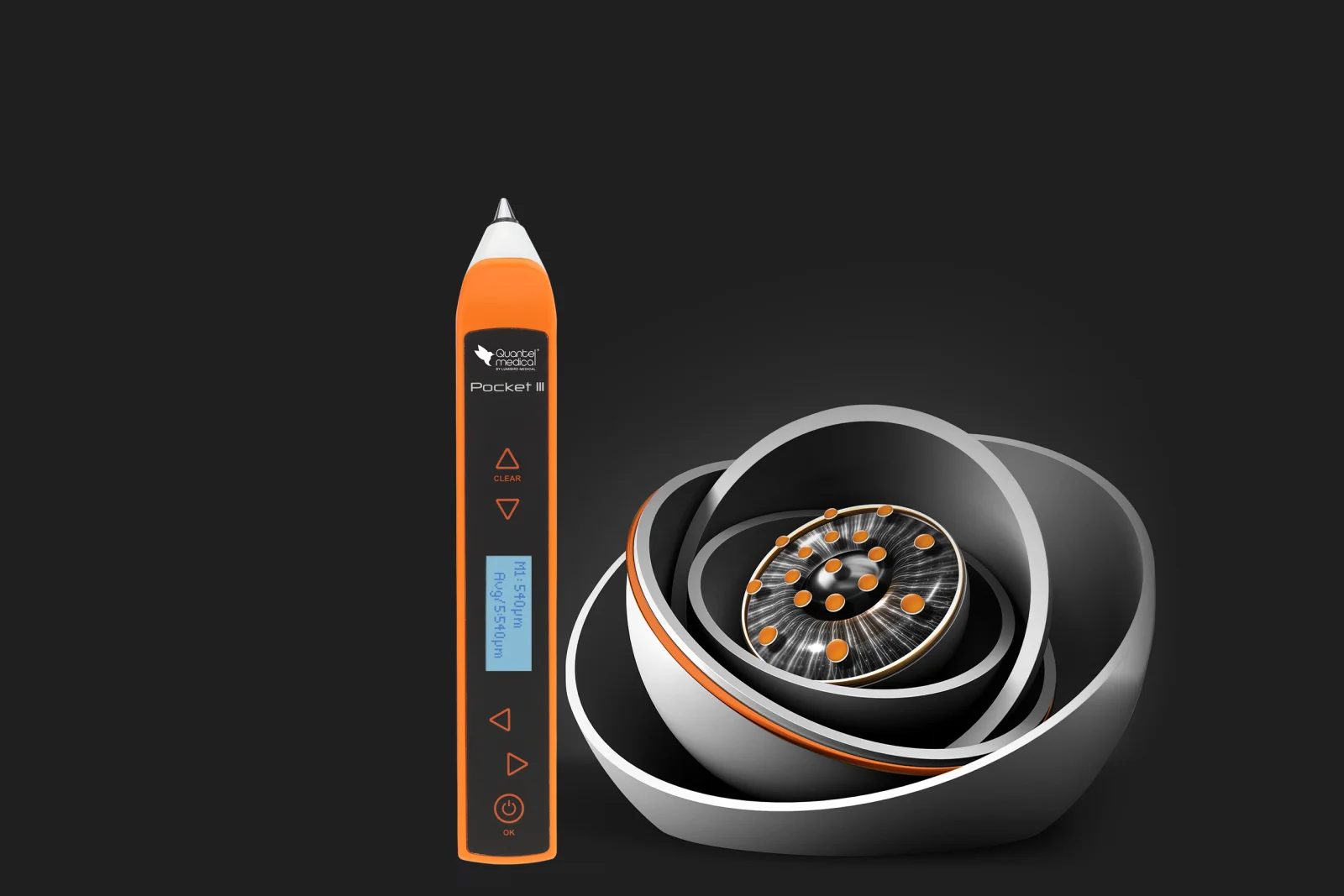
The POCKET III is a handheld contact ultrasound pachymeter that measures corneal thickness at the point of care. It uses a 30 MHz transducer with a 150 to 1200 µm range, and has built-in IOP correction tables. It supports glaucoma follow-up, refractive screening and corneal monitoring.
Core Technology Specifications
Imaging Platform: A-scan ultrasound pachymetry (contact, pulse-echo)
System Type: Handheld, connected, ambidextrous pachymeter
Probe Frequency: 30 MHz
Measurement Range: 150 to 1200 µm
Accuracy: ± 5 µm
Modes: Central measurement and corneal mapping
Weight: 60 g
Regulatory Status: CE marked and FDA 510(k)
How It Measures
The probe contains a piezoelectric transducer that emits a short ultrasound pulse at 30 MHz. The pulse produces two main echoes: one from the anterior corneal surface (tear film and epithelium) and one from the posterior surface (endothelium and aqueous humor interface). The device measures the time between the two echoes and converts it to thickness using an assumed speed of sound in corneal tissue. The distance is halved, because the pulse travels to the interface and back.
Frequency affects resolution. A higher frequency means a shorter wavelength and better axial resolution, which suits a thin structure like the cornea. The trade-off is reduced tissue penetration, which does not matter at this depth.
Anatomical Context
The cornea has five layers: epithelium, Bowman's layer, stroma, Descemet's membrane and endothelium. The stroma makes up about 90% of the total thickness. Ultrasound pachymetry measures the total thickness from the anterior surface to the endothelial interface. The central cornea averages roughly 540 µm in healthy adults, and it is thinner centrally than peripherally.
Why Corneal Thickness Matters
- Tonometry: Applanation readings depend on corneal thickness. A thick cornea can overestimate IOP, and a thin cornea can underestimate it
- Glaucoma risk: Central corneal thickness is a recognized factor in assessing risk in ocular hypertension
- Refractive surgery: Laser ablation removes stromal tissue, so residual thickness must be sufficient
- Corneal disease: Thickness changes help in following keratoconus, ectasia and edema
Built-in IOP Correction
The device includes correction tables relating IOP to corneal thickness (Ehlers, Doughty and Dresdner). These tables are population-based approximations, so corrected values should be interpreted alongside the rest of the clinical picture.
Measurement Technique
- Anesthesia: Topical anesthetic is required, as it is a contact measurement
- Alignment: Hold the probe perpendicular to the central cornea. An oblique angle lengthens the ultrasound path and overestimates thickness
- Central mode: Gives a central thickness reading
- Mapping mode: Takes readings at several points for a regional picture
- Display: Shows each reading and the running average
- Verification: The holder has a test disk for checking the probe's measurement and for practicing perpendicular positioning
Factors Affecting Accuracy
- Probe angle and decentration: The most common sources of operator error
- Pressure on the cornea: Excess pressure can compress the tissue and underestimate thickness
- Tear film and epithelial changes: These can alter the anterior echo
- Hydration state: Edematous corneas change the actual acoustic properties of the tissue
- Post-surgical corneas: The assumed speed of sound may not match altered stromal structure, so values should be interpreted with caution
- Repeatability: Averaging several readings improves reliability
Hardware and Ergonomics
- Weight: 60 g, described as the lightest connected handheld pachymeter on the market
- Handling: Ambidextrous pen-style body that supports perpendicular positioning
- Interface: Navigation arrows (including CLEAR), an OK/power button and a small LCD
- Power: Rechargeable lithium-ion battery
Connectivity and Data
- Transfer: Bluetooth or USB-C
- Software: POCKET III Manager (Windows 10 compatible)
- Memory: 10 patients (20 eyes maximum)
Hygiene
The probe tip rests in a dedicated holder with a disinfection cup. This supports infection control between patients.
Clinical Applications
- Glaucoma and ocular hypertension assessment
- Pre-refractive surgery screening
- Keratoconus and ectasia follow-up
- Corneal edema monitoring (range extends to 1200 µm)
- Thin cornea assessment (range starts at 150 µm)
- Portable measurement in clinics, screenings or bedside settings
Who Can Use It
Ophthalmologists, optometrists and orthoptists working in glaucoma, cornea, refractive and general practice.
Comparison with POCKET II
| Feature | POCKET II | POCKET III |
|---|---|---|
| Range | 200–999 µm | 150–1200 µm |
| Accuracy | ± 5 µm | ± 5 µm |
| Modes | Central and mapping | Central and mapping |
Strengths and Limits
Strengths: Portable, fast, simple to use, and an extended range that covers thin and edematous corneas.
Limits: Contact technique requiring anesthesia, operator-dependent alignment, and point measurements rather than the full thickness maps given by topography or tomography systems.
Limitations of Available Specifications
Note: The assumed speed of sound, resolution, probe tip diameter, battery runtime, dimensions, display specifications and operating conditions were not available in the documentation reviewed. Contact Quantel Medical or an authorized distributor for the complete datasheet and pricing. This content supports clinical judgement and does not replace it.
Visreed