

OPMI LUMERA 700 - ZEISS
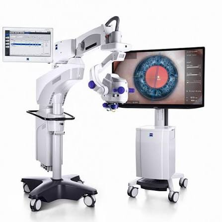
From the manufacturer
Zeiss: official product page
zeiss.com · opens in a new tab
Abstract
The OPMI LUMERA 700 is ZEISS's ophthalmic surgical microscope, mainly for cataract and anterior segment surgery. It combines Stereo Coaxial Illumination (SCI) for a bright, stable red reflex, fully apochromatic optics with a motorized 1:6 zoom, motorized focus and XY positioning, and an integrated slit illuminator. It can be extended with the RESCAN 700 intraoperative OCT, the CALLISTO eye guidance system and TRENION 3D HD visualization, and it is part of the ZEISS Cataract Suite.
Core Technology Specifications
Instrument Type: Ophthalmic surgical microscope, floorstand or ceiling-mounted
Illumination: Stereo Coaxial Illumination (SCI); xenon (Superlux Eye) or LED fiber-optic light source
Optics: Fully apochromatic optics with motorized zoom, DeepView depth of field management
Magnification: 0.4x to 2.4x (zoom ratio 1:6) with 10x wide-angle eyepieces (12.5x optional)
Objective Lens: f = 200 mm standard; f = 175 mm or 225 mm available
Focus Range: 70 mm, electric focus
XY Coupling: Maximum travel 61 x 61 mm with automatic centering
Slit Illuminator: Integrated, widths 0.2, 2, 3 and 4 mm, length 12 mm, with left-right movement
User Profiles: Settings for up to 20 users, with a programmable wireless foot pedal
Integrated Display: Integrated 22-inch monitor (1,680 x 1,050) for viewing in the operating room, HD video recording
Anatomical Context: Why the Red Reflex Matters
In cataract surgery, the surgeon looks through the pupil at the lens. Light that passes through the pupil, reflects from the retina and returns through the eye gives the red reflex, a glow behind the lens that outlines the capsule and the cataractous material. A bright, even red reflex helps the surgeon see the anterior capsule during capsulorhexis and the lens fragments during phacoemulsification, especially in dense or white cataracts.
The same illumination also matters in corneal and other anterior segment surgery, where contrast and depth of field help in handling thin tissue. Red reflex quality therefore depends on how the light is delivered into the eye, which is the core design question for a cataract microscope.
How the System Works
- Stereo Coaxial Illumination: SCI provides coaxial and full-field illumination from the same optical axis as the observation path. ZEISS describes the result as a stable, high-contrast red reflex that stays bright even when the eye or the microscope moves
- Light sources: The xenon source is a Superlux Eye short-arc lamp with a HaMode filter and a manual backup lamp. The LED source has a near-daylight color temperature and a stated lifetime of 50,000 hours at 50% intensity. Both support an optional blue-blocking filter and an optional fluorescence filter
- Optics: The apochromatic design corrects color errors to give a whiter and more natural image, and DeepView manages depth of field
- Motorization: Electric focus and XY coupling, with automatic centering, let the surgeon control position without moving the hands. The spring arm carries up to 9 kg of additional accessories
- Slit illumination: The integrated slit illuminator lets the surgeon check the anterior chamber and cornea during surgery without changing instruments
Optional Modules and Integration
- RESCAN 700: Spectral-domain intraoperative OCT built into the microscope, with a wavelength of 840 nm, 27,000 A-scans per second and an axial resolution of 5.5 µm in tissue. Scan depth can be set to 2.9 or 5.8 mm, the scan can be rotated through 360°, and both live and capture modes are available. An XY tracker keeps the scan location during the procedure
- CALLISTO eye: A guidance system with a touchscreen panel PC and software (version 3.7 listed). It offers markerless toric IOL alignment (Z ALIGN), corneal curvature estimation (K TRACK) and LRI planning, so manual corneal marking is not needed
- RESIGHT 700: A motorized lens revolver for posterior segment viewing, supplied with 60 D macular and 128 D wide-field lenses
- TRENION 3D HD: A 3D HD stereoscopic visualization system, so that students and staff can follow the surgery in three dimensions outside the eyepieces
- Documentation: Recording of the surgical view and data by USB, with compatibility with the ZEISS FORUM data management system
ZEISS Cataract Suite
The LUMERA 700 is one component of the ZEISS Cataract Suite. The suite links biometry data from the IOLMaster, planning parameters, markerless alignment with CALLISTO eye and video documentation. The aim is a connected workflow from the pre-operative measurement to the operating room.
Reported Benefits
- Time saving: ZEISS states that CALLISTO eye saves about 6 minutes per cataract procedure by removing the manual marking steps
- Surgeon testimonials: The brochure quotes surgeon testimonials, for example 99% of toric IOL patients within ±0.50 D of residual cylinder, and 4.2 minutes less graft manipulation time in corneal surgery
- Interpretation: These are manufacturer figures and individual surgeon reports, not results of a controlled trial
Clinical Applications
- Cataract surgery, including dense and white cataracts, with a strong red reflex
- Toric IOL implantation with markerless alignment
- Corneal and anterior segment surgery, with intraoperative OCT for depth information
- Posterior segment viewing with the RESIGHT 700 lens revolver
- Teaching and documentation through the 3D visualization and video recording
Who Can Use It
Cataract, corneal and anterior segment surgeons in the operating room. Surgical assistants and residents can follow the case on the integrated monitor or the 3D display, and the independent zoom on the assistant's microscope supports teaching. Orthoptists and optometrists are not users of this device, but they work with the pre-operative measurements that feed into it.
Comparison of Light Sources
| Feature | Xenon (Superlux Eye) | LED |
|---|---|---|
| Color | White, natural, high contrast | Near-daylight color temperature |
| Lifetime | Lamp, with a backup lamp | 50,000 h at 50% intensity |
| Filters | HaMode, blue-blocking, fluorescence option | HaMode, blue-blocking, fluorescence option |
| Illumination mode | SCI | SCI |
Strengths and Limits
Strengths: A bright and stable red reflex, apochromatic optics, motorized controls with user profiles, a modular route from a standard microscope to intraoperative OCT and toric guidance, and a link to the biometry workflow.
Limits: Intraoperative OCT, CALLISTO eye and 3D visualization are separate modules, so the full configuration is a larger investment. It is a fixed operating room installation, and its weight, dimensions and power requirements were not available. The benefit claims come from ZEISS and surgeon testimonials.
About ZEISS
ZEISS Meditec is the medical technology business of Carl Zeiss. In ophthalmology it covers surgical microscopes, biometry such as the IOLMaster, diagnostic imaging and refractive lasers. The LUMERA 700 belongs to its surgical microscope family.
Limitations of Available Specifications
Note: The sources reviewed were the ZEISS product page, an ophthalmology portal and a distributor brochure. The weight, overall dimensions, power supply, regulatory status by country, the exact light output and pricing were not available. Some pages list slightly different component details, such as the integrated monitor size, so please check the current ZEISS data sheet for your configuration. This content supports clinical judgement and does not replace it.