

MeiboVue - VisuScience
Abstract
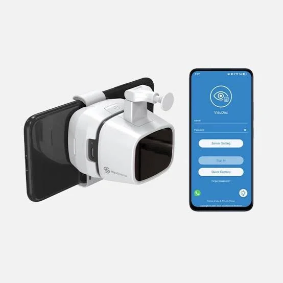
MeiboVue (model VMC-100) is a handheld meibographer that clips onto a smartphone. It images the meibomian glands of the eyelids with LED illumination and sends the images over Wi-Fi to the VisuDoc app and to a computer. It is designed for fast screening of meibomian gland dysfunction (MGD) and dry eye assessment, in the exam room or away from an instrument table.
Core Technology Specifications
Imaging Platform: Meibography with a dedicated camera head and LED illumination
System Type: Portable, smartphone-based meibographer (the device is the camera, the phone is the screen)
Sensor: 5 MP (VMC-100); a 10 MP version (VMC-200) is listed in the manual
Working Distance: 35 mm
Image Format: JPEG
Image Resolution: 2592 x 1944 (manual) or 2560 x 1600 (specification page)
Preview: Live preview at 1920 x 1080, 25 fps (manual)
Data Transfer: Wi-Fi (5 GHz bands listed in the FCC statement)
App: VisuDoc, iOS and Android, with PC software for Windows and Mac
Power: Rechargeable Li-ion battery, 3.7 V / 2500 mAh, about 3 hours of working time; input DC 5 V / 1 A
Dimensions and Weight: 67 x 60 x 60 mm (L x W x H), 350 g
Regulatory Status: The manufacturer states that it is FDA approved (please verify the exact clearance for your region)
Anatomical Context: the Meibomian Glands
The meibomian glands are sebaceous glands embedded in the tarsal plates of the upper and lower eyelids. Their orifices open along the eyelid margin. They secrete meibum, an oily mixture that forms the outer lipid layer of the tear film and slows tear evaporation.
In MGD, the glands become obstructed or atrophic. The most common form is obstructive, with thickened secretion and blocked orifices. This reduces the lipid layer and leads to evaporative dry eye. On meibography, gland drop-out, shortening and distortion are visible, which is why imaging helps in diagnosis and follow-up.
How the System Works
- Imaging: The camera head is held in front of the everted eyelid. LED illumination makes the glands visible, and the live image shows on the smartphone screen
- Phone as display: The smartphone sits on a clip on the device. It acts as the screen for preview and capture, while the camera head does the imaging
- Wireless link: The camera sends the live video and captured images to the phone over Wi-Fi
- Voice-activated capture: The practitioner can capture images with a voice command, which leaves both hands free to hold the lid and the camera
- Data flow: Images move from the phone to a PC through VisuDoc and can then be uploaded to an EMR or EHR system
Workflow
- Speed: The manufacturer states that the glands of one eyelid can be captured in about 30 seconds
- Eyelid eversion: The LidVue eyelid everter is supplied in the kit to help expose the tarsal conjunctiva
- Technician use: The live preview helps the technician focus and capture a good image for the doctor to review later
- Data storage: The VisuDoc PC software is installed locally on the computer, and the manufacturer states that patient data stays on the user's device
What Is in the Kit
- Meibomian gland imaging camera
- LidVue eyelid everter
- Power adaptor and USB-C charging cable
- Software USB disc and instructions for use
- Carry box
- Slit lamp attachment
- Android phone (listed in the kit specification)
Clinical Applications
- Screening for meibomian gland dysfunction
- Dry eye pre-test imaging and documentation
- Follow-up of treatment response, by comparing images over time
- Patient education, by showing the glands on the screen
- Satellite clinics, bedside examination and community screening events
Who Can Use It
Ophthalmologists and optometrists in dry eye and cornea practice. Technicians and orthoptists can capture the images in a pre-test area, and the clinician then reviews them. The manufacturer designed it for all types of optometry and ophthalmology clinics.
Comparison with Desktop Meibography
| Feature | Desktop meibography | MeiboVue |
|---|---|---|
| Setup | Instrument table | Handheld, no table |
| Mobility | Fixed in one room | Portable, carry box |
| Capture | Operator at the instrument | Smartphone and voice command |
| Transfer | Integrated software | Wi-Fi to VisuDoc, then PC and EMR |
| Slit lamp use | Not applicable | Slit lamp attachment included |
Strengths and Limits
Strengths: Portable, fast capture, hands-free voice command, wireless transfer to the EMR workflow, and a slit lamp attachment for flexibility.
Limits: The image quality depends on correct eyelid eversion and focus. At 350 g it is heavier than a pen-style device, and it needs a Wi-Fi link to the phone. Automated gland analysis or grading was not described in the documentation reviewed, so interpretation is done by the clinician.
About VisuScience
VisuScience Meditech is the manufacturer. Its portfolio of ophthalmic accessories includes MeiboVue, QuikVue (a smartphone adaptor for anterior segment imaging), iFlo, iExpressor and LidVue.
Limitations of Available Specifications
Note: The LED wavelength (infrared or visible), field of view, depth of field, any built-in gland analysis or grading tools, and the exact regulatory clearance number were not available in the documentation reviewed. Resolution values differ between the manual and the specification page. Contact VisuScience or an authorized distributor for the full datasheet and pricing. This content supports clinical judgement and does not replace it.
Visreed