

GAIA - Topcon
From the manufacturer
Topcon: official product page
topconhealthcare.eu · opens in a new tab
Abstract
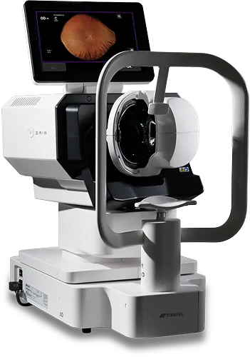
GAIA is a fully automated, non-contact, non-mydriatic ultra-widefield retinal imager from Topcon Healthcare. It uses slit-scanning illumination with RGB LED light sources to produce true-color images. Topcon advertises a 209° field of view in a single capture, but its own footnote says this is an estimate derived from a measured angular field of 150°. It can image through pupils of 2.0 mm or more, and it integrates with IMAGEnet 7 and DICOM systems. It received a CE mark in 2026, and Topcon says it is not approved or offered in every market.
Status and Availability
- Europe: The CE mark was announced in an article dated 21 July 2026. Topcon planned commercial availability in the EU in the weeks that followed, with a launch at ESCRS 2026 in London (11 to 15 September)
- Other markets: The Topcon Japan page states that GAIA is not approved or offered in every market, and that availability varies by country
- Not stated: The pages reviewed list no FDA clearance, price or country list. Check your local situation with Topcon
Core Technology Specifications
Instrument Type: Ultra-widefield retinal slit scanner, fully automated, non-contact
Illumination: Structured slit light with a high-speed rolling shutter, which the manufacturer says blocks scattered light; red, green and blue LED sources
Image Depth: 24-bit true color
Field of View: 209° advertised (an estimate); measured angular field 150° with a pupil of 2.5 mm or more, and 120° with a pupil of 2.0 mm
Resolution: Color imaging resolving power of 20 lp/mm or more
Pupil Requirement: 2.0 mm or more, confirmed with model eyes; no pharmaceutical dilation
Dioptric Range: −20 D to +20 D
Modes: Color and infrared (the infrared entry carries an asterisk that is not explained)
Automation: Auto-alignment, auto-focus and auto-shoot
Connectivity: USB flash drive, shared network folders, DICOM, DICOM server storage, and integration with IMAGEnet 7
Dimensions and Weight: 334 to 646 (W) x 549 to 573 (D) x 672 to 702 mm (H), as listed in ranges; 31 kg
Understanding the 209° Figure
Fundus camera field of view can be described as an angle measured from the pupil, or as an angle at the eye's center that covers the retinal surface. The two are not the same, and the second gives a larger number for the same image. Topcon's footnote says that the 209° figure is an estimate converted from a 150° visual angle, using a relationship published by Yao et al. in Biomedical Optics Express (2021).
| Value | Condition | Note |
|---|---|---|
| 209° | Single capture, advertised | Estimate derived from 150° |
| 150° | Pupil 2.5 mm or more | Measured angular field of view |
| 120° | Pupil 2.0 mm | Measured angular field of view |
When comparing GAIA with other ultra-widefield devices, make sure the same angle convention is used. A 209° figure can look larger than a competitor's number that is not converted.
Anatomical Context: Why the Peripheral Retina Matters
A standard fundus camera images the posterior pole, the macula and the optic disc. Many retinal diseases also start or show signs in the periphery, such as peripheral lesions in diabetic retinopathy, retinal tears and degenerations, and vascular changes. Ultra-widefield imaging aims to show more of this peripheral retina in a single picture, so that fewer images or manual steering are needed.
Imaging the periphery through a small pupil is difficult, because the pupil and the optics of the eye limit the light that enters and leaves. This is why the pupil size requirement and the field of view depend on each other, as the table above shows.
How the System Works
- Slit scanning: The structured slit of light scans across the retina while a high-speed rolling shutter reads only the illuminated area. Topcon says this effectively blocks scattered light, which would otherwise lower contrast
- True color: Red, green and blue LEDs illuminate the retina, and the page describes a full-color image of 24-bit depth
- Automation: Auto-alignment, auto-focus and auto-shoot make the imaging fully automated, which Topcon says gives patient comfort and an easy workflow
- Data handling: Images can be exported through USB, shared folders or DICOM, and stored on a DICOM server. IMAGEnet 7 is described as creating a unified patient record across modalities
- Not stated: The page does not state the scan time, the wavelengths of the LEDs or of the infrared source, or any AI function
Clinical Applications
- Imaging of the peripheral retina in a single capture
- Documentation of retinal disease, for example diabetic retinopathy; the Topcon page shows examples before and after additional laser treatment
- Imaging through small pupils without dilation
- Topcon's president of eye care business states that a wider view of the peripheral retina may support early detection and management of retinal disease
- No intended use statement, clinical study or performance data was found in the sources reviewed
Strengths and Limits
Strengths: A very wide advertised field of view, true color imaging, a small minimum pupil, fully automated operation, non-contact and non-mydriatic use, and links to DICOM and IMAGEnet 7.
Limits: The 209° figure is an estimate and the measured field is 150° (120° with a 2.0 mm pupil). The page lists only color and infrared modes, so autofluorescence and angiography are not stated. It weighs 31 kg and is not approved in every market. No independent clinical evidence was found.
About Topcon Healthcare
Topcon Healthcare is the eye care and healthcare technology business of Topcon, with imaging systems such as OCT and fundus cameras and the IMAGEnet software platform. GAIA was shown together with the Maestro3 series and MYAH PRO at ESCRS 2026.
Limitations of Available Specifications
Note: The specifications come mainly from Topcon's Japan product page, and the availability and CE mark details come from Topcon's European pages and trade news that repeat the company's statements. The scan time, LED wavelengths, infrared details, power requirements, autofluorescence or red-free modes, AI features, FDA status, price and any clinical studies were not available. The background on peripheral retinal disease and field of view conventions is standard clinical knowledge and was not taken from the sources. Please check the current Topcon documentation for your country. This content supports clinical judgement and does not replace it.
Sources
- Topcon Healthcare Japan: GAIA product page: https://topconhealthcare.jp/ultra-wide-field-retinal-slit-scanner/gaia/
- Topcon Healthcare Europe: Introducing GAIA: https://topconhealthcare.eu/en_EU/gaia
- Topcon Healthcare Europe: GAIA and Maestro3 at ESCRS 2026 (news): https://topconhealthcare.eu/en_EU/news/topcon-healthcare-unveils-gaia-and-maestro3-series-powered-by-imagenet-r-7-myah-pro-clinical-decision-support-solutions-for-glaucoma-and-a-new-patient-guided-refraction-system-at-the-escrs-2026-in-london
- AOP Optometry Today: CE mark approval for Topcon's Gaia (21 July 2026): https://www.aop.org.uk/ot/news/2026/07/21/ce-mark-approval-for-topcons-gaia
- Eyewire+: Topcon Receives CE Mark for GAIA: https://eyewire.news/news/topcon-receives-ce-mark-for-gaia-ultra-widefield-retinal-imaging-system