

FUNDO - OPTOPOL
From the manufacturer
Optopol: official product page
optopol.com · opens in a new tab
Abstract
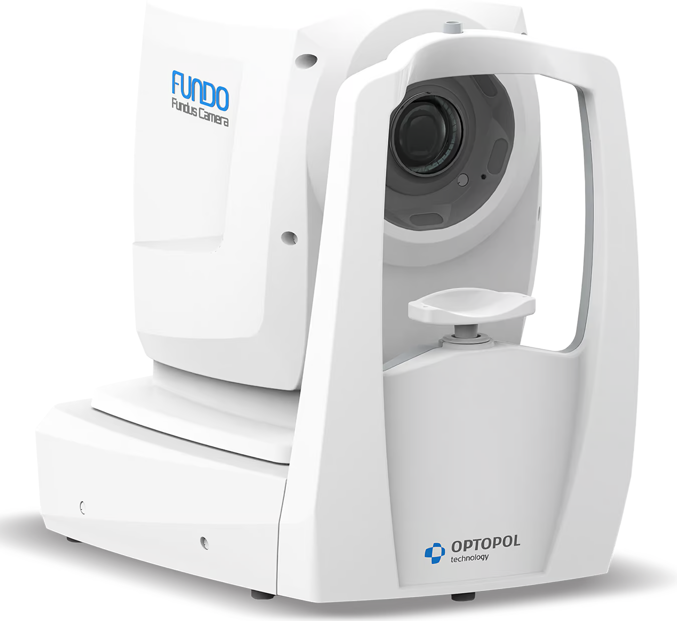
FUNDO is a non-mydriatic automatic fundus camera from OPTOPOL Technology. It has a 20.3 MP CMOS sensor, a 90° field of view with a standard pupil, a mosaic mode up to 198°, and a minimum pupil size of 3.3 mm. It detects the pupil, aligns, focuses and adjusts exposure automatically. Besides color and infrared fundus imaging, it lists anterior segment imaging, meibography and optional autofluorescence, FA and ICGA modes. Images are managed in OPTOPOL's REVO application. At the time of writing (November 2026), the OPTOPOL website lists FUNDO as coming soon.
Status of This Product
The only source for the specifications below is the manufacturer's product page, which marks FUNDO as coming soon. The OPTOPOL news page shows no launch date, announcement or availability. No independent source, clinical study or regulatory information was found. Treat all figures as manufacturer pre-launch specifications that may change.
Core Technology Specifications
Instrument Type: Non-mydriatic automatic fundus camera, tabletop
Sensor: 20.3 MP CMOS
Field of View: 90° (60°) ±5% with a standard pupil; 67° (45°) ±5% with a small pupil; optional mosaic mode 198° (132°) ±5%. The page does not explain the figures in parentheses
Minimum Pupil Size: 3.3 mm
Working Distance: 31 mm
Focus Range: −25 D to +25 D
Capture Modes: Color fundus and anterior segment; infrared (IR) fundus and anterior segment; meibography; optional FAF Green, FAF Blue, FA and ICGA
Flash, Gain and Exposure: Automatic or manual; intensity levels High, Normal and Low
Alignment: Fully automatic, automatic or manual; automatic pupil detection, alignment, focus and exposure adjustment
Fixation Targets: Internal OLED display with adjustable shape and position, steady or blinking with adjustable rate; external target available
Software: REVO application; DICOM, network connectivity and export to a chosen location
Dimensions and Weight: 490 x 350 x 530 mm (L x W x H); 19 kg
Power: 100 to 240 V, 50/60 Hz; 90 to 110 VA
Anatomical Context: What a Fundus Camera Images
The fundus is the interior back surface of the eye. It includes the retina, the macula and fovea, the optic disc, the retinal vessels and the periphery. A fundus camera photographs it through the pupil, using the same optical path to light the retina and to collect the image. The pupil therefore limits both the light that gets in and the area that can be seen.
In a non-mydriatic camera, the examiner images through a natural, undilated pupil, usually in a darkened room with infrared light for alignment, so the pupil is not constricted by visible light before the flash. This is why the minimum pupil size matters. A wider field of view shows more of the retina, which helps to document the periphery as well as the macula and optic disc.
Field of View and Pupil Size
| Condition | Field of view (as listed) | Note |
|---|---|---|
| Standard pupil | 90° (60°) ±5% | Single image |
| Small pupil | 67° (45°) ±5% | Minimum pupil 3.3 mm |
| Mosaic mode (optional) | 198° (132°) ±5% | Composite of several images |
How the System Works
- Automation: The camera detects the pupil and aligns itself, then adjusts focus and exposure. The operator can choose fully automatic, automatic or manual alignment
- Fixation: The internal OLED display provides a fixation target whose shape, position and blink rate can be adjusted, so the patient can be guided to look in the direction needed for each retinal area. An external target is also available
- Light control: Flash, gain and exposure can be automatic or manual, at three intensity levels
- Focus: The focus range of −25 D to +25 D covers a wide range of refractive errors without a separate correction
- Eye tracking: The page lists no eye tracking function. Whether the camera compensates for eye movement during capture is not stated
Imaging Modes
- Color: Standard color photography of the fundus and of the anterior segment
- Infrared: Infrared imaging of the fundus and of the anterior segment
- Fundus autofluorescence (optional): Optional FAF Green and FAF Blue modes, which show the autofluorescence of the retinal pigment epithelium
- FA and ICGA (optional): Optional fluorescein angiography and indocyanine green angiography, for vascular imaging
- Meibography: Meibography is listed, for imaging the meibomian glands in dry eye work
- Red-free: A Digital Red-Free filter appears as an analysis filter, not as a capture mode
Software and Connectivity
- REVO: Images are acquired and managed in the REVO application
- Data transfer: DICOM support, network connectivity and export to a chosen location
- OPTOPOL ecosystem: FUNDO integrates with OPTOPOL OCT and visual field systems on a common patient database with REVO and REVO 2
- AI and comparison: The page mentions AI-highlighted anomaly review and chronological comparison to monitor progression. It gives no details of the AI function, its validation or its regulatory status
Clinical Applications
- Retinal diagnostics, including the macula, optic disc, retinal vessels and periphery
- Imaging without routine dilation
- Anterior segment documentation
- Meibography for meibomian gland dysfunction
- Comparison of images over time to follow progression
- Angiography and autofluorescence, with the optional modules
Strengths and Limits
Strengths: High sensor resolution, a wide field of view and a mosaic mode, automatic alignment and exposure, an internal fixation display, a wide range of imaging modes in one device, and connection to the OPTOPOL OCT and visual field database.
Limits: It is not yet on the market according to the source, and there is no price, regulatory status or independent evidence. Angiography and autofluorescence are optional. The page does not list eye tracking. A weight of 19 kg makes it a fixed tabletop unit.
About OPTOPOL
OPTOPOL Technology is a manufacturer of ophthalmic diagnostic devices, including OCT and fundus camera systems such as the REVO line. The sources reviewed state no relationship with other ophthalmic companies.
Limitations of Available Specifications
Note: All product figures come from a single manufacturer page for a product listed as coming soon. The meaning of the numbers in parentheses next to the fields of view, the flash type, whether the camera has eye tracking, the AI function, regulatory status, availability and pricing were not available. The anatomy and non-mydriatic imaging background is standard clinical knowledge and was not taken from the source. Search results for other OPTOPOL cameras (REVO FC) describe different devices and were not used. Please check the current OPTOPOL documentation. This content supports clinical judgement and does not replace it.
Sources
- OPTOPOL: FUNDO product page (listed as coming soon): https://optopol.com/products/fundo/
- OPTOPOL: News page (checked for a FUNDO announcement; none found): https://optopol.com/news/